Anatomy Muscles Pelvis / 79 Pelvic Girdle Vectors Free Royalty Free Pelvic Girdle Vector Images Depositphotos. They have several functions, including helping to support the pelvic organs. Some of the major pelvic muscles are as follows. It attaches to the walls of the lesser pelvis, separating the in this article, we shall look at the anatomy of the muscles that make up the inferior lining of the cavity; The pelvic floor muscles provide foundational support for the intestines and bladder. The right and left hip bones also converge anteriorly to attach to each other.
Rectus femoris muscle, one of the quadriceps muscles on the front of your thigh. The muscles of the femoral region of the lower limb are divided into three compartments. Ligaments of the pelvis and hip. The adductor muscle group, also known as the groin muscles, is a group located on the medial side of the thigh. The gluteal muscles are a group of three muscles named the gluteus maximus, the gluteus medius, and the gluteus minimus.
The Pubococcygeal Muscle Pc Muscle And Attachments Yoganatomy from cdn-aolkg.nitrocdn.com Anatomy of pelvic and acetabular muscles. The stability and flexibility of the hip joint is provided by two structures: The levator ani muscles are the largest group of muscles in the pelvis. These muscles have attachments to the pelvis as follows: Ligaments, tendons, and muscles play an important role in the function of the hip. During the closed chain phase, which is when the foot is in contact with the ground, the muscle externally rotates the femur on th … anatomy, bony pelvis and lower limb, popliteus muscle review. The levator ani muscles consist of three. The pelvic diaphragm is the third deepest layer of the pelvic floor which puts it at the very center of all the other muscles.
To support the abdominal and pelvic viscera
It's supplied by ventral rami of first and 2nd sacral nerves (s1, s2). The pelvic girdle (hip girdle) is formed by a single bone, the hip bone or coxal bone (coxal = hip), which serves as the attachment point for each lower limb. The pelvic floor muscles are comprised of 3 layers and have a complex relationship with the surrounding bony pelvis, fascia, ligaments and nerves. Attached to the pelvis are muscles of the buttocks, the lower back, and the thighs. 12 photos of the muscle anatomy pelvis. The levator ani is a broad sheet of muscle. The main function of the pelvic floor muscles are: The muscles of the pelvic floor are collectively referred to as the levator ani and coccygeus muscles. Cross sectional muscle anatomy of pelvis, ct pelvic muscle anatomy, mri pelvic muscle anatomy, pelvic muscle anatomy, pelvic muscular anatomy, human muscles, cross sectional muscle anatomy of pelvis, ct pelvic muscle anatomy, mri pelvic muscle anatomy, pelvic muscle anatomy, pelvic muscular anatomy. The medial surface provides attachment for both transverse perinei, obturator internus and externus, piriformis, coccygeus and levator ani muscles. The anterior compartment includes pectineus, iliopsoas, psoas minor, iliacus. Several muscles around the pelvis take part in movements of the thigh. It attaches to the walls of the lesser pelvis, separating the in this article, we shall look at the anatomy of the muscles that make up the inferior lining of the cavity;
The thigh bone or femur and the pelvis join to form the hip joint. The stability and flexibility of the hip joint is provided by two structures: Each compartment is separated from the others by an intermuscular septum that runs from the fascia lata to the linea aspera of the femur. During the closed chain phase, which is when the foot is in contact with the ground, the muscle externally rotates the femur on th … anatomy, bony pelvis and lower limb, popliteus muscle review. Each hip bone, in turn, is firmly joined to the axial skeleton via its attachment to the sacrum of the vertebral column.
Budget Pelvis Model With Organs And Pelvic Floor Muscles Anatomystuff Co Uk from www.anatomystuff.co.uk The main function of the pelvic floor muscles are: (1) the obturator internus and the piriformis, which are muscles of the lower extremity, and will be described with these (pages 476 and 477); Created by physicians for you to help you understand the pelvic floor. The right and left hip bones also converge anteriorly to attach to each other. The pelvic girdle (hip girdle) is formed by a single bone, the hip bone or coxal bone (coxal = hip), which serves as the attachment point for each lower limb. The ischium provides numerous points of attachment for pelvic and lower limb muscles. This mri male pelvis axial cross sectional anatomy tool is absolutely free to use. Use the mouse scroll wheel to move the images up and down alternatively use the tiny arrows (>>) on both side of the image to move the images.>>) on both side of the image to move the images.
The bones of the pelvis are held together by a large number of ligaments and muscles.
Some of the major pelvic muscles are as follows. The pelvic diaphragm is the third deepest layer of the pelvic floor which puts it at the very center of all the other muscles. (2) the levator ani and the coccygeus, which together form the pelvic diaphragm and are associated with the pelvic viscera. Muscles play an important role in the. The floor of the pelvis is made up of the muscles of the pelvis, which support its. The gluteal muscles are a group of three muscles named the gluteus maximus, the gluteus medius, and the gluteus minimus. (1) the obturator internus and the piriformis, which are muscles of the lower extremity, and will be described with these (pages 476 and 477); Anatomy of pelvic and acetabular muscles. 12 photos of the muscle anatomy pelvis. The muscles found within the true pelvis include the piriformis muscles, obturator internus muscles, and muscles of the pelvic diaphragm. The stability and flexibility of the hip joint is provided by two structures: The pelvis is the lower portion of the trunk, located between the abdomen and the lower limbs. Use the mouse scroll wheel to move the images up and down alternatively use the tiny arrows (>>) on both side of the image to move the images.>>) on both side of the image to move the images.
Whenever someone talks about the pelvic floor in general, they are probably talking about these 5 muscles: These muscles have attachments to the pelvis as follows: The labeled structures are (excluding the correct side): Ligaments of the pelvis and hip. The muscles within the pelvis may be divided into two groups:
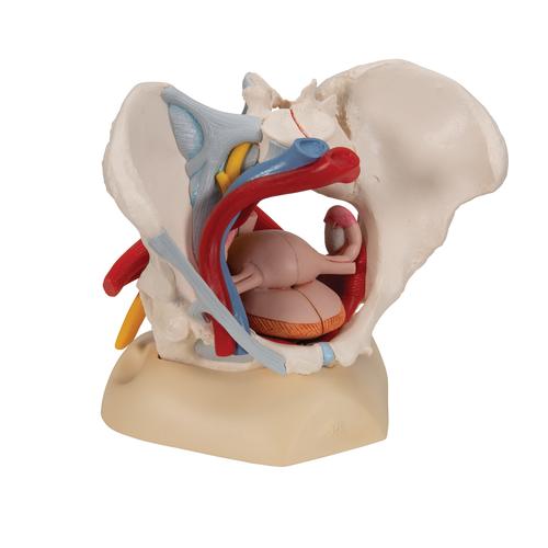
Anatomical Teaching Models Plastic Human Pelvic Models Female Pelvis With Ligaments Vessels Nerves Pelvic Floor Muscles And Organs from www.3bscientific.com Anatomy of pelvic and acetabular muscles. They have several functions, including helping to support the pelvic organs. Muscles that attach from the pelvis to the trunk and cross the lumbosacral joint muscles that attach from the pelvis to the thigh/leg and cross the hip joint pelvic floor muscles that are located wholly within the pelvis The muscles found within the true pelvis include the piriformis muscles, obturator internus muscles, and muscles of the pelvic diaphragm. The pelvic girdle (hip girdle) is formed by a single bone, the hip bone or coxal bone (coxal = hip), which serves as the attachment point for each lower limb. Ligaments of the pelvis and hip. The gluteal muscles are a group of three muscles named the gluteus maximus, the gluteus medius, and the gluteus minimus. Each hip bone, in turn, is firmly joined to the axial skeleton via its attachment to the sacrum of the vertebral column.
See more ideas about anatomy, thoracic, basic image.
The thigh bone or femur and the pelvis join to form the hip joint. These muscles origin in continuity from the body of the pubis, along a tendinous arch over the obturator internus fascia, and the ischial spine. The pelvic floor muscles provide foundational support for the intestines and bladder. Ƒ pelvic floor dysfunction is common and. The pelvic floor muscles are comprised of 3 layers and have a complex relationship with the surrounding bony pelvis, fascia, ligaments and nerves. 12 photos of the muscle anatomy pelvis. The labeled structures are (excluding the correct side): Some of the major pelvic muscles are as follows. It's supplied by ventral rami of first and 2nd sacral nerves (s1, s2). The pelvis's frame is made up of the bones of the pelvis, which connect the axial skeleton to the femurs, and therefore acts in weight bearing of the upper body. Muscles that attach from the pelvis to the trunk and cross the lumbosacral joint muscles that attach from the pelvis to the thigh/leg and cross the hip joint pelvic floor muscles that are located wholly within the pelvis Rectus femoris muscle, one of the quadriceps muscles on the front of your thigh. These muscles have attachments to the pelvis as follows:
Share :
Post a Comment
for "Anatomy Muscles Pelvis / 79 Pelvic Girdle Vectors Free Royalty Free Pelvic Girdle Vector Images Depositphotos"


Post a Comment for "Anatomy Muscles Pelvis / 79 Pelvic Girdle Vectors Free Royalty Free Pelvic Girdle Vector Images Depositphotos"